The patient arrives via private vehicle, limping heavily, with a visibly deformed knee. “My kneecap popped out,” she says. “Can you just pop it back in?” As field providers, we face this scenario regularly — and the evidence increasingly supports our ability to reduce patellar dislocations safely in the prehospital setting. But what exactly are we managing, and what does the science tell us about outcomes?
What is a patellar dislocation?
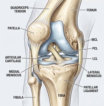
The patella — your kneecap — sits in a groove at the lower end of the femur called the trochlear groove. This groove normally keeps the patella centered as the knee bends and straightens. A patellar dislocation occurs when the patella displaces out of this groove, almost always laterally (outward). The kneecap literally shifts out of its normal position, disrupting the biomechanics of the entire knee joint.
Anatomy matters here. The patella is held in place by soft-tissue structures — the quadriceps muscle and tendon above, the patellar ligament below, and medial and lateral retinaculum on either side. When these structures fail — usually from a twisting injury to a planted foot, a direct blow, or a sudden direction change — the patella can slip out of its groove. While some dislocations spontaneously reduce (the kneecap pops back in on its own), many remain displaced and require manual intervention.
Imaging the dislocation
X-ray confirmation is the gold standard. On a lateral view, a dislocated patella appears displaced anteriorly and laterally from the femoral condyles. The axial (sunrise) view clearly shows the patella sitting outside the trochlear groove. The anteroposterior (AP) view adds information about associated fractures, which occur in roughly 5–10% of acute patellar dislocations.
Associated injuries frequently accompany patellar dislocations:
- Osteochondral fractures (bone and cartilage fragments)
- Medial retinaculum tears
- Quadriceps muscle contusions
- Effusion (fluid buildup) within the joint
- Ligamentous injuries to the MCL or ACL
These associated injuries are precisely why hospital imaging remains mandatory — even after a successful field reduction.
Typical field presentation
The presentation is usually unmistakable. Patients with acute patellar dislocations typically report:
Chief complaint: sudden, severe knee pain, often preceded by an audible or palpable “pop.”
History of injury:
- Non-contact mechanism: deceleration, pivoting, cutting movements
- Contact: direct blow to the medial knee
- Twisting injury with a planted foot
- Common in athletes during sports, but also occurs in falls or simple missteps
Visible findings:
- The kneecap visibly displaced laterally — patients can usually point to where it is
- Severe, rapidly developing swelling (within minutes)
- Skin tension over the displaced patella
- Significant bruising, typically developing within hours
Patient behavior:
- Unwillingness to bear weight on the affected limb
- Knee held in slight flexion (patient resists full extension)
- Severe anxiety — patients are frightened by the visible deformity
- Urgent, justified requests for pain relief
Transient reductions matter. Some patients will have already spontaneously reduced by the time you arrive. Don’t assume the injury was minor — these patients still have significant soft-tissue damage and require full evaluation and imaging.
Paramedic assessment
1. Scene safety and primary survey
As with any trauma, rule out life threats first. Patellar dislocations are typically isolated injuries, but confirm the mechanism didn’t involve higher-energy trauma (high-speed falls, motor vehicle collisions) that might suggest additional injuries.
2. Focused history
- Exact moment of injury onset
- Mechanism (pivoting, fall, direct blow, deceleration)
- Did the kneecap relocate on its own? When?
- Prior dislocations? (Recurrent dislocations suggest predisposing anatomic factors)
- Pain severity and location
- Any “giving way” or instability before this event?
3. Neurovascular assessment (critical)
This is non-negotiable. A dislocated patella can compromise blood flow to the lower leg, particularly if swelling is significant or there’s concurrent injury. Document:
- Distal pulses: palpate posterior tibial and dorsalis pedis pulses bilaterally; compare to the unaffected leg
- Capillary refill: should be <2 seconds in the toes
- Skin color and temperature: a pale, cool lower leg suggests compromised perfusion
- Motor function: can the patient wiggle toes? Plantarflex? Dorsiflex?
- Sensory function: gross sensation intact in toes and foot?
Red flags — immediate physician evaluation, don’t delay transport
- Absent distal pulses
- Capillary refill >2 seconds
- Pale or cyanotic foot
- Motor or sensory deficits
- Significant deformity making assessment unreliable
4. Physical examination of the knee
- Inspect: swelling and obvious lateral displacement of the patella
- Palpate: effusion (ballottement test if swollen), joint-line tenderness, crepitus
- Range of motion: don’t force it. Assess passive range gently — most patients can’t tolerate active extension initially
5. Special tests
If the patella is still displaced, a gentle patellar apprehension test — light lateral pressure on the medial border with the knee flexed ~20–30 degrees — helps confirm lateral displacement.
How to reduce: the technique
The evidence-based prehospital reduction is straightforward, safe, and highly effective. It requires two providers and takes roughly 30 seconds to 2 minutes.
- Patient positioning. Place the patient supine on the stretcher; the affected leg relaxed and slightly flexed. Explain the procedure to reduce anxiety.
- Pre-reduction confirmation. Verify distal pulses and neurovascular status one final time. Confirm you’ve ruled out associated fractures as much as possible. If you suspect a proximal tibia or distal femur fracture, do NOT attempt reduction — transport immediately.
- Provider positioning. Provider A handles the leg from beside the affected limb; Provider B positions on the opposite side with access to the lateral knee.
- Flex the hip. Provider A gently flexes the patient’s hip (aim for ~45 degrees), supporting the lower leg and foot. This relaxes the quadriceps, reducing tension and making reduction easier.
- Extend the knee. Provider A slowly and steadily extends the knee with gentle, controlled pressure — never forceful. Some dislocations reduce spontaneously during this phase.
- Medial manual pressure. If it hasn’t reduced, Provider B applies steady, firm pressure to the lateral edge of the patella, pushing it medially back into the groove. You’ll often feel or hear a “clunk.” Pressure should be firm and steady — not painful or forceful. Most reductions occur within 30 seconds.
- Confirm reduction. The patella should be centered in the groove, the patient typically feels immediate, dramatic pain relief, and the knee should extend more fully.
- Post-reduction care. Immobilize the knee in full or near-full extension, ice if available, elevate the limb, and re-assess distal neurovascular status. Transport for definitive evaluation and imaging.
Analgesia considerations
The evidence here is clear: analgesia is often unnecessary for the reduction itself. Anxiety relief, a clear explanation, and the dramatic pain relief immediately after a successful reduction mean many patients tolerate the procedure without additional medication. That said, protocols vary:
- Some systems use nitrous oxide or intranasal fentanyl pre-reduction
- Some use procedural sedation for a highly anxious patient or after failed initial attempts
- Gentle technique and adequate explanation reduce the need for significant pain management
- Post-reduction pain control is reasonable and important for comfort during transport
Pros and cons of field reduction
Advantages
- Immediate pain relief. Pain is often 8–10/10; successful reduction relieves it within seconds — this alone justifies the procedure.
- Reduced opioid requirement. Pre-reduction analgesia needs are minimal in many cases, and post-reduction comfort is achieved with basic analgesics.
- Improved distal perfusion. Reducing the dislocation lets swelling redistribute and may improve perfusion. Profound vascular compromise is rare — but why risk it?
- Faster transport. A reduced, immobilized knee means less scene time and often a non-emergent transport.
- Improved patient experience. Eliminating the visible deformity removes a major psychological stressor.
- Safety record. Multiple studies and protocols show prehospital reduction is safe when performed per protocol, with minimal adverse events.
Risks and complications
- Associated fractures. The biggest risk is reducing in the presence of a proximal tibia or distal femur fracture — hence careful history and palpation first. If fracture is suspected, immobilize and transport without reducing.
- Rare neurovascular injury. Forceful reduction can injure soft tissue; gentle, evidence-supported technique minimizes this.
- Inadequate pain control. If the patient can’t tolerate the procedure, use analgesia/sedation if protocol allows, or transport unreduced — ED physicians reduce it routinely.
- Spontaneous re-dislocation. The patella can re-dislocate if the knee moves — which is why immediate immobilization matters. You can re-attempt reduction or transport.
- Inadequate assessment. Missing associated injuries is the biggest pitfall. Maintain high suspicion, palpate carefully, and don’t hesitate to transport without reducing if something seems wrong.
The evidence
Support for prehospital patellar dislocation reduction has grown substantially over the past decade. A protocol study in Prehospital Emergency Care (2019) demonstrated that a prehospital reduction protocol was safe and effective, with high success rates and minimal adverse events. Multiple EMS systems have since incorporated the skill — New York implemented it at the BLS level after demonstrating patient benefit — and complications from gentle, protocol-driven reduction remain remarkably rare. The improvement in patient comfort and the reduction in opioid requirements are well documented.
The bottom line
Patellar dislocations are common, painful, and visually dramatic. The evidence increasingly supports prehospital reduction when performed per protocol by trained providers with appropriate patient selection. The key elements:
- Careful assessment for associated injuries, especially proximal tibia/femur fractures
- Gentle technique — not forceful manipulation
- Appropriate analgesia as needed, but often less than expected
- Immediate immobilization after reduction
- Hospital transport for imaging and evaluation of associated injuries
If your system doesn’t yet have a prehospital patellar dislocation reduction protocol, the evidence supports advocating for one. Your patients will appreciate the dramatic pain relief, and you’ll be providing evidence-based, safe prehospital care that genuinely improves outcomes.
References
- Implementation of a Prehospital Patella Dislocation Reduction Protocol — Prehospital Emergency Care (2019)
- EMS Evaluation and Management of Limb-Threatening Knee Injuries — JEMS
- How to Reduce a Lateral Patellar Dislocation — Merck Manual Professional Edition
- Dislocated Kneecap — Cleveland Clinic
- Patella Dislocation — StatPearls (NIH)
- Irreducible Lateral Patellar Dislocation: Case Report and Literature Review — PMC