Patient Scenario
A wet December morning a 68-year-old female steps onto her front porch to grab the morning newspaper. She doesn’t realize its iced over, she slips, extends her outstretched arm to break her fall, and instead fractures her wrist. On arrival she is seated inside her warm home, guarding her right forearm against her stomach with her left hand. On first glance the wrist has a class “fork” presentation, and you are suspicious of a Colle’s Fracture. Swelling is already challenging the integrity of the two rings on her hand. Other than embarrassment and pain, she is alert and oriented without distracting or additional injuries.
This call will feel like a speed bump in a career of emergency response, and yet it is one of the most common injury patterns. The care in the next 20 minutes matters more than most EMTs realize.
This short form essay will focus on upper extremity fractures, immobilization and treatment for the BLS provider.
Upper Extremity Focus
In a national sample of more than 87 million Americans, researchers counted 590,193 upper extremity fractures in one year, an annual incidence of 67.6 per 10,000 persons.1 Distal radius and ulna fractures were the most common, followed by hand fractures, proximal humerus fractures and clavicle fractures. Distal radius fractures followed a bimodal pattern, with the highest rates in patients under 18 and over 65.1 A Swedish national registry of more than 23,000 adult distal radius fractures found that a simple fall caused about three quarters of them, and the most typical patient was an older woman injured at home.2 Our patient on the front steps is the textbook case.
Most of these injuries are not life threatening, and that is exactly why they get treated casually. The arm is packed with structures that do not forgive mistakes: the subclavian vessels behind the clavicle, the radial nerve wrapped around the humeral shaft, the brachial artery crossing the front of the elbow, and the median nerve passing through the wrist. The fracture is the obvious problem. The neurovascular structures beside it are where the real risk lives.
Anatomy
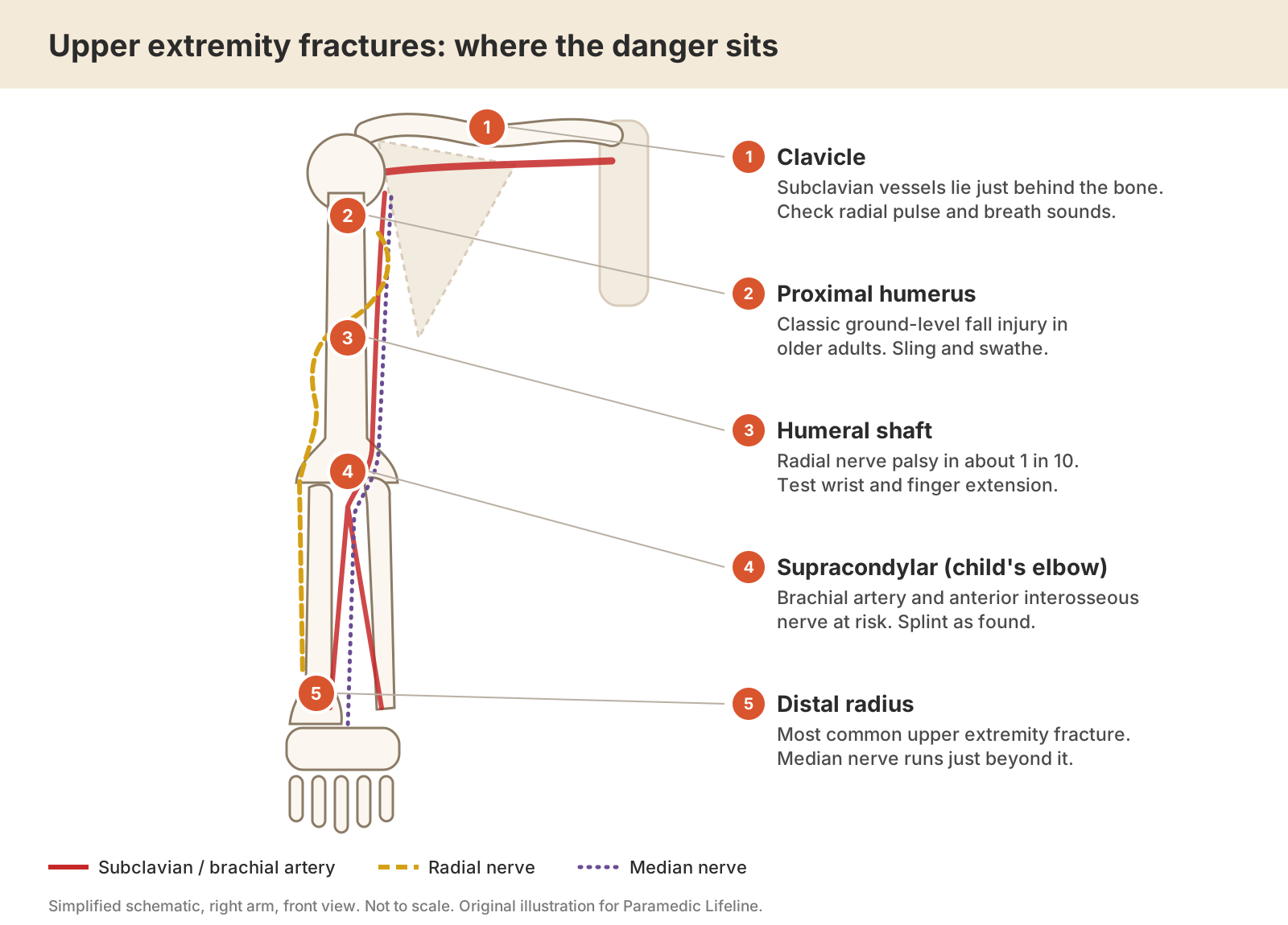
Five fracture sites and the structures beside them. Original schematic for Paramedic Lifeline, compiled from the sources cited in the text.
Clavicle
Most clavicle fractures heal without trouble, but the subclavian artery and vein run directly behind the bone. Clavicle fractures are uncomfortable and impact sleeping, self-care and other activities of daily living. A 2024 review estimated subclavian vessel injury at roughly 0.3 to 3 percent of clavicle fractures, more likely after high-energy mechanisms and with displaced or comminuted fractures. The authors also warned that the obvious pain of the fracture can delay recognition of the vascular injury.3
Proximal humerus and humeral shaft
Proximal humerus fractures are among the most common upper extremity fractures overall.1 Shaft fractures put the radial nerve at risk as it spirals along the back of the bone. In a systematic review of 1,758 patients with closed humeral shaft fractures, primary radial nerve palsy occurred in 10 percent, and more than 90 percent of those palsies recovered without further intervention.4 An earlier review of 4,517 fractures reported a similar prevalence of 11.8 percent, with fractures of the middle and distal shaft carrying the highest risk.5 The field finding is wrist drop: the patient cannot extend the wrist or fingers.
Elbow
Supracondylar humerus fractures in pediatric patients is the elbow injury that worries orthopedic surgeons most. Up to 12 percent carry an associated nerve injury.6 In the common extension-type fracture, the anterior interosseous nerve, a motor branch of the median nerve, is the nerve most often injured.7 Because that branch has no skin territory of its own, a quick "can you feel this" check will miss it. The brachial artery crosses the front of the elbow, and a systematic review of trauma-related forearm compartment syndrome found supracondylar fractures to be the most common fracture behind it.8
Forearm and wrist
Distal radius fractures typically follow a fall on an outstretched hand. The median nerve runs through the carpal tunnel just beyond the fracture site, where swelling can compress it. Both-bone forearm fractures and distal radius fractures follow supracondylar fractures as the leading fracture causes of forearm compartment syndrome.8
Typical field presentation
Patients with arm fractures usually tell you where the problem is, and our visual “look test” assessment often identifies the injury. Patients will exhibit guarding, and resist movement. Look for deformity, swelling, bruising, point tenderness, shortening or angulation. Crepitus may be felt during handling; note it, but never go looking for it. Three patterns are worth recognizing on sight: the dorsal dinner fork deformity of the wrist, the older adult holding the shoulder stiffly against the chest after a fall, and the child who will not move a swollen elbow.
Two details deserve special attention. The first is mechanism. A ground-level fall in an older adult raises its own questions about why the patient fell, whether the head was struck and whether an anticoagulant is on board. Is this a slip and fall, a syncope, an arrythmia or a CVA?
A high-energy mechanism moves the priority from the arm to the whole patient. A patient who crashed his motorcycle and is complaining of wrist pain, may easily have other injuries. This bolsters the already important step of the head-to-toe primary assessment.
BLS assessment, step by step
- Life threats come before limbs. Major extremity bleeding is controlled with direct pressure or a tourniquet. In a multisystem trauma patient, a deformed arm is a distraction; manage the airway, breathing and circulation, start transport, and splint enroute if there is time. Remove clothing from the injured limb, then take off rings, watches and bracelets before swelling makes it impossible.
- Compare distal pulses, capillary refill, skin color and temperature with the uninjured side. North Carolina's statewide splinting procedure is explicit: assess and document pulses, sensation and motor function before the splint goes on, and again after.9
- Radial nerve: "thumbs up" with the wrist extended, and sensation in the web space between thumb and index finger on the back of the hand. Median and anterior interosseous nerves: make a round "OK" sign with thumb and index finger, and test sensation at the tip of the index finger. Ulnar nerve: spread the fingers apart, and test sensation on the little finger. A child who cannot make a round O despite normal sensation may have an anterior interosseous nerve injury.
- Use a standard pain scale and record it before and after each intervention, as the NASEMSO model pain protocol recommends.10
- Document what you find in plain terms. A deficit present at the scene is part of the injury. A documented baseline lets the receiving team recognize a new deficit if one appears later, and that distinction drives decisions about imaging and specialty consultation.
Core splinting principles
- Immobilize the joint above and the joint below a long bone fracture. For a joint injury, immobilize the bone above and the bone below.
- Pad generously, filling voids and protecting bony prominences. For forearm and wrist injuries, place a roll of gauze in the palm to hold the hand in a position of function.
- Splint injuries involving the shoulder, elbow or wrist in the position found. Never force a deformed elbow straight.
- Secure the splint above and below the injury, not directly over it.9
- Reassess circulation, motor and sensation after splinting and during transport. If any of the three has worsened, loosen or remove the splint and reassess.9
- Support the splinted arm with a sling and swathe so its weight is not hanging from the fracture site.
Injury by injury
The table below condenses the approach for each region. Local protocol governs the specific devices and techniques your agency uses.
Injury | Typical mechanism | What to check | BLS splinting approach |
|---|---|---|---|
Clavicle | Fall onto the shoulder or outstretched arm; direct blow | Radial pulse, hand color and warmth; breath sounds and chest wall when the mechanism is high energy | Sling and swathe in a position of comfort |
Proximal humerus / shoulder | Ground-level fall in an older adult | Sensation over the lateral shoulder; distal pulses; head strike and anticoagulant use | Sling and swathe; pad the axilla; a shoulder held away from the body is splinted as found with pillows or blankets |
Humeral shaft | Fall, direct blow, twisting injury | Wrist and finger extension; sensation in the dorsal first web space | Padded rigid splint along the arm, then sling and swathe so the arm does not hang |
Elbow, including the pediatric supracondylar fracture | Fall on an outstretched hand, often a child from playground equipment | Radial pulse and capillary refill; OK sign; finger spread; pain that keeps climbing | Splint in the position found; never force it straight; early notification if the hand is pulseless |
Forearm shaft (radius and ulna) | Direct blow, fall, sports | Forearm firmness; pain with passive stretch of the fingers | Rigid splint from above the elbow to the hand, elbow flexed about 90 degrees, then sling |
Distal radius / wrist | Fall on an outstretched hand, older adult | Index fingertip sensation and OK sign (median nerve); capillary refill | Padded rigid or vacuum splint from the elbow to the fingertips; gauze roll in the palm; sling |
Hand and fingers | Punch, crush, ball sports | Rotational deformity, capillary refill, rings | Remove rings; splint in position of function or buddy tape per protocol |
BLS quick reference for upper extremity fractures.
The pulseless arm
Most prehospital protocols allow for reduction or correction of angulation when the distal circulation is impaired.9 In practice, for an angulated long bone shaft fracture, that usually means one gentle attempt to bring the limb toward normal alignment with steady in-line traction, stopping if pain or resistance increases, and only where local protocol and medical direction allow it. Suspected elbow and shoulder joint injuries are not manipulated. They are splinted as found, the receiving facility is notified early, and transport is expedited.
In dual response agency models (BLS, ALS) there is often an associated procedural sedation protocol that should be considered if time and circumstances allow.
The pediatric elbow deserves particular respect. Surgeons describe a "pink, pulseless hand" after supracondylar fracture, in which the hand looks perfused, but no radial pulse can be felt. That appearance can be falsely reassuring. In a long-term follow-up series of 26 children referred to a specialist unit with this presentation, 23 already had established ischemic contracture of the forearm and hand.11 A child with an elbow injury and no radial pulse, or with pain that keeps climbing, is a time-sensitive patient who should go to a facility capable of definitive pediatric orthopedic care according to local destination guidelines.
Ice, Elevation and POC
Position of comfort, cold packs and splinting are the first-line nonpharmacologic measures for traumatic pain in the NASEMSO model protocol.10 Wrap cold packs so they never sit directly on skin, and elevate the splinted arm when the patient's condition allows. Be honest about the evidence for ice, though. A systematic review of randomized trials found few studies of ice in closed soft-tissue injury and no evidence for an optimal mode or duration,12 and an emergency medicine review concluded that evidence that cryotherapy improves clinical outcome was insufficient.13 Ice is cheap, safe when wrapped and often welcome. It is a comfort measure, not a treatment that changes healing.
Pain Management
Splinting is analgesia, and good positioning is analgesia, nonetheless some patients need additional pain management. The 2019 National EMS Scope of Practice Model added oral over-the-counter analgesics to the EMT level,14 as allowed by protocol and EMS Medical Director. Oral analgesics often have a longer absorption and time to therapeutic threshold and for cases likely to require surgery be cautious of oral ingestion that may delay surgery due to anesthesiology risk parameters. Older adults deserve particular attention here: the NAEMSP 2025 position statement on geriatric trauma calls for prompt pain evaluation and timely analgesic intervention in this population.15
Open Fractures
Control bleeding, cover the wound with a sterile dressing (moistened with sterile saline where protocol directs), do not push protruding bone back under the skin, and splint as found. Record the time of injury. That time matters because infection risk climbs with delay. In a study of 137 patients with severe (type III) open tibia fractures, antibiotics given more than 66 minutes after injury independently predicted deep infection.16 A 2024 joint position statement from the American College of Surgeons Committee on Trauma, the Orthopaedic Trauma Association, ACEP, NAEMSP and NAEMT supports giving antibiotics for suspected open fractures as soon as possible, including before hospital arrival.17 Antibiotics sit outside BLS scope in most systems, but are covered here conceptually due to national guidance. The BLS job is to recognize the open fracture, report it early and give the ALS crew or emergency department the information they need to start the clock sooner.
Compartment Syndrome
Compartment syndrome usually develops over hours, so it is most likely to surface on long transports, interfacility transfers or patients found long after injury. It can also develop after casting or immobilization with air splints if swelling internally is compression/restricting against the immobilization device. In conscious patients, pain out of proportion to the injury and pain on passive stretch of the muscles are the most useful clinical observations; changes in sensation, weakness and pallor tend to come late.18 For the forearm, gently extending the fingers is the passive stretch test. A splint or bandage applied too tightly can cause or worsen the problem, so loosen it, reassess and report what you found. Splinted and immobilized limbs should be reassessed along with Q5/Q10 vital signs enroute via checking for distal pulse and sensation, and pain score.
Evidence
Extremity splinting rests largely on anatomy, physiology and expert consensus rather than randomized trials, and most prehospital fracture research has focused on the femur. The stronger evidence comes from the hospital literature on complications, and it supports the specific vigilance described above. Radial nerve palsy is common with humeral shaft fractures and usually recovers.4 Nerve palsies after pediatric supracondylar fractures also tend to recover; in a review of 510 such palsies, 376 resolved without investigation or intervention.6 A small but real minority of upper extremity fractures threaten the limb through vascular injury, ischemia or compartment syndrome.3,8,11
That pattern defines the value of good BLS care. Most patients will do well regardless of what we do, provided we do no harm. The crew's real contribution is a careful baseline exam, a splint that protects without compressing, adequate pain control and early recognition of the few fractures that are limb threatening.
Bottom line
Throughout your EMS career you will encounter many fractures, and as your experience grows you will learn from those patient care episodes and your peers what works best. Most EMS rigs contain various options for immobilization – padded short and long boards, air splints, slings, backboards, and traction devices. Each scenario requires some creativity in determining the best approach. Join our discussions on our webpage or on the substack and share your tips and pearls.
End Notes
- Karl JW, Olson PR, Rosenwasser MP. The epidemiology of upper extremity fractures in the United States, 2009. J Orthop Trauma. 2015;29(8):e242-e244. doi:10.1097/BOT.0000000000000312
- Rundgren J, Bojan A, Mellstrand Navarro C, Enocson A. Epidemiology, classification, treatment and mortality of distal radius fractures in adults: an observational study of 23,394 fractures from the national Swedish fracture register. BMC Musculoskelet Disord. 2020;21:88. doi:10.1186/s12891-020-3097-8
- Chalidis B, Davitis V, Papadopoulos P, Pitsilos C. Subclavian vessels injury: an underestimated complication of clavicular fractures. World J Crit Care Med. 2024;13(4):98579. doi:10.5492/wjccm.v13.i4.98579
- Hendrickx LAM, Hilgersom NFJ, Alkaduhimi H, Doornberg JN, van den Bekerom MPJ. Radial nerve palsy associated with closed humeral shaft fractures: a systematic review of 1758 patients. Arch Orthop Trauma Surg. 2021;141(4):561-568. doi:10.1007/s00402-020-03446-y
- Shao YC, Harwood P, Grotz MRW, et al. Radial nerve palsy associated with fractures of the shaft of the humerus: a systematic review. J Bone Joint Surg Br. 2005;87(12):1647-1652. doi:10.1302/0301-620X.87B12.16132
- Graff C, Dounas GD, Todd MRLC, Sung J, Kumawat M. Management of traumatic nerve palsies in paediatric supracondylar humerus fractures: a systematic review. Children (Basel). 2023;10(12):1862. doi:10.3390/children10121862
- Babal JC, Mehlman CT, Klein G. Nerve injuries associated with pediatric supracondylar humeral fractures: a meta-analysis. J Pediatr Orthop. 2010;30(3):253-263.
- Khoshhal KI, Alsaygh EF, Alsaedi OF, Alshahir AA, Alzahim AF, Al Fehaid MS. Etiology of trauma-related acute compartment syndrome of the forearm: a systematic review. J Orthop Surg Res. 2022;17:342. doi:10.1186/s13018-022-03234-x
- North Carolina College of Emergency Physicians. Standards Procedure (Skill): Splinting (Procedure 44). North Carolina Office of EMS.
- National Association of State EMS Officials. Model EMS Protocol for Prehospital Pain Management. May 2021.
- Blakey CM, Biant LC, Birch R. Ischaemia and the pink, pulseless hand complicating supracondylar fractures of the humerus in childhood: long-term follow-up. J Bone Joint Surg Br. 2009;91-B(11):1487-1492. doi:10.1302/0301-620X.91B11.22170
- Bleakley C, McDonough S, MacAuley D. The use of ice in the treatment of acute soft-tissue injury: a systematic review of randomized controlled trials. Am J Sports Med. 2004;32(1):251-261. doi:10.1177/0363546503260757
- Collins NC. Is ice right? Does cryotherapy improve outcome for acute soft tissue injury? Emerg Med J. 2008;25(2):65-68.
- National Highway Traffic Safety Administration and National Association of State EMS Officials. National EMS Scope of Practice Model 2019 (DOT HS 812 666). Washington, DC: NHTSA; 2019.
- Haussner WK, Breyre AM, Bascombe K, et al. Prehospital trauma compendium: management of geriatric trauma patients. A position statement and resource document of NAEMSP. Prehosp Emerg Care. 2025.
- Lack WD, Karunakar MA, Angerame MR, Seymour RB, Sims S, Kellam JF, Bosse MJ. Type III open tibia fractures: immediate antibiotic prophylaxis minimizes infection. J Orthop Trauma. 2015;29(1):1-6. doi:10.1097/BOT.0000000000000262
- American College of Surgeons Committee on Trauma, Orthopaedic Trauma Association, American College of Emergency Physicians, National Association of EMS Physicians, National Association of Emergency Medical Technicians. Prehospital antibiotic administration for suspected open fractures: joint position statement. 2024.
- Ali P, Santy-Tomlinson J, Watson R. Assessment and diagnosis of acute limb compartment syndrome: a systematic literature review. Int J Orthop Trauma Nurs. 2014;18(4):180-190. doi:10.1016/j.ijotn.2014.01.002
Image credits
Figure 1 and Figure 2 are original digital illustrations created for Paramedic Lifeline. They do not depict real patients and contain no third-party copyrighted material. The quick reference table was compiled by the author from the sources listed above.