Recognizing and treating hyperkalemia before the lab result exists
Steve Rinaldi, MBA, NRP | paramediclifeline.com
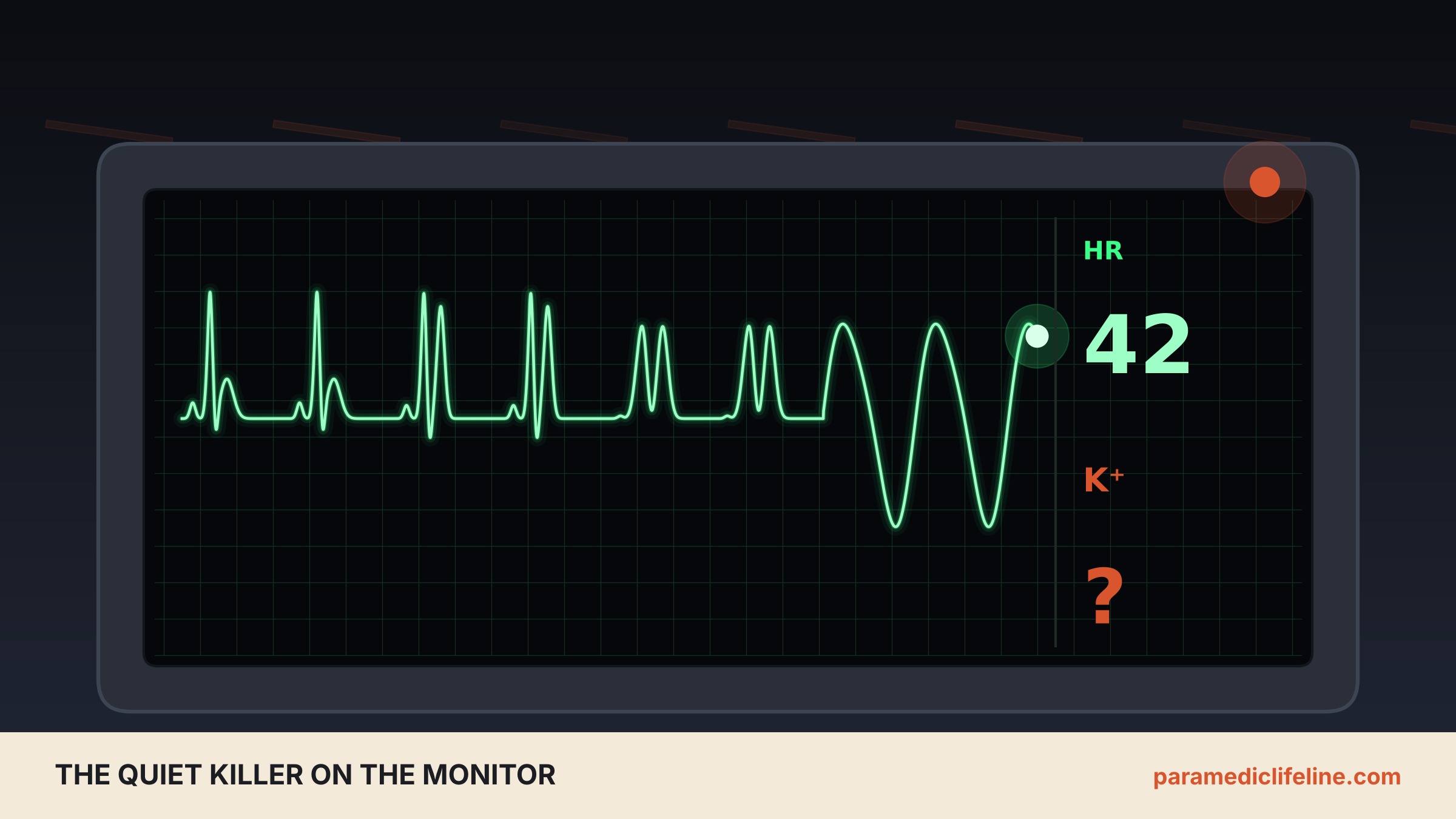
Illustration: Paramedic Lifeline. Original computer-generated artwork created for this article; not a patient image or a real recording.
Opening Scenario
It is a Sunday afternoon, and you are dispatched to a 64-year-old man who is "too weak to stand." His wife says he skipped his Saturday dialysis because he felt like he was coming down with something. You already start to be concerned, because you understand how physiologically significant a missed dialysis visit can be. He is alert, pale and frustrated that his legs "won't work." His blood pressure is 92/58. The monitor shows a heart rate in the low 40s with no obvious P waves and a QRS that looks a little too wide. The T waves are not especially tall.
So far you have not identified any “priority symptoms”; all the pertinent negatives are just that, negative. Despite this, our patient may be minutes from a fatal arrythmia. You have down a metabolic panel, but the serum potassium score, that will confirm, is still an hour or more in the future after the hospital laboratory runs the sample. The next 10 minutes depends upon what you see on the monitor and how you piece together the history.
Hyperkalemia
Potassium is the dominant cation inside cells, and the steep gradient between intracellular and extracellular potassium is what lets nerves fire and heart muscle depolarize and repolarize in an orderly way. In case you were wondering, I will save you the search, the most abundant intracellular anion is phosphate. When extracellular potassium climbs, that gradient narrows. Repolarization speeds up, which produces the narrow, peaked T wave, and conduction through the atria, the AV node and the ventricles slows, which shows up as a longer PR interval and a wider QRS.2 Push far enough and the QRS and T wave merge into a sine wave that precedes ventricular fibrillation, PEA or asystole.5